6 December 2021
What is it like to have a lumbar puncture?
What is it like to have a lumbar puncture? A woman who underwent a lumbar puncture during her diagnosis for MS (multiple sclerosis) shares her personal account of the process.
A lumbar puncture is a diagnostic test for multiple sclerosis that involves removing and analysing a sample of cerebrospinal fluid (CSF), the fluid that surrounds the brain and spinal cord within the skull and backbone. It is sometimes referred to as a spinal tap.
A lumbar puncture takes about half an hour under a local anaesthetic. The lumbar region is the section of the backbone between the lowest ribs and the pelvis. A thin, flexible needle is inserted into the base of the spine above the pelvis and a quantity of cerebrospinal fluid is drawn off.
Having a lumbar puncture can be uncomfortable or unsettling. The drop in pressure in the cerebrospinal fluid caused by the removal of a sample can produce a splitting headache for some people. This usually lasts for no more than 24 hours but for some can persist for longer. To reduce the risk of headaches, you should try to lie flat for at least six hours after the procedure and drink plenty of water.
There is a lower risk of headaches and infections following lumbar puncture if the clinician uses a special atraumatic needle, sometimes called a Sprotte needle. These are not always commonly used, but they are recommended for use by the BMJ and you can ask if you are concerned.
The fluid that is drawn off in a lumbar puncture is analysed to look for a number of different things.
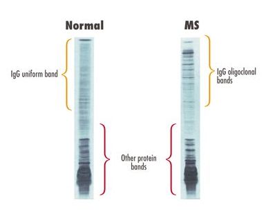
The test that shows the presence of antibodies is called electrophoresis. A sample of fluid is placed on a gel and voltage is applied. This causes antibodies of the same size to bunch together, forming visible 'bands'.
One band (monoclonal) in the cerebrospinal fluid is normal. The term 'oligoclonal bands' refers to the presence of two or more bands and shows the presence of disease activity. Whilst this doesn't necessarily mean that someone has MS, about 80-95% of people with MS do have oligoclonal banding in their cerebrospinal fluid.
